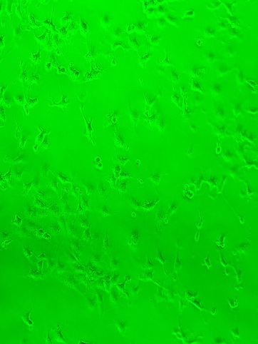
These are my cells. There are many like them, but these are mine. What are shown are cos7 cells, cells that are derived from monkey kidney tissue. Early in my internship experience, my mentor was kind enough to give me these cells to work with throughout my journey. These cells played a huge role in my internship. With them, I learned skills such as cell lysing, serum starving, and most importantly, cell transfection. Transfection is when you introduce nucleic acid, such as DNA or RNA, from another source into a cell. As part of my project, I would be transfecting these cos7 cells with green fluorescent protein (GFP). GFP is the protein found in marine organisms, like jellyfish, that cause fluorescence under a certain wavelength. During experiments, you can link GFP to other plasmids, so that you can see if your cells were properly transfected, merely by checking to see if your cells are glowing. GFP can also be put in tumours so that you can visualize where the tumour detaches, and migrates to. GFP was actually in-part discovered by a professor at UCSD (Roger Y. Tsien). While I did not get the chance to meet him during my internship, I took advantage of his discovery throughout the completion of my project. When I arrived at internship, I had no understanding about how I would be able to transfect my cells, but soon I would learn.
Lesson 1: Labeling. The first thing my mentor taught me through this process was that I must always label everything— absolutely everything. While this seemed to be such a simple concept to me, I did not realize at the time how important of a lesson this was. Eventually, I began to lyse my cells, which is breaking down the cells to get the contents you want. I added the buffer needed for lysing, spun down my cells, then was about to remove the contents I wanted into another set of tubes. I then realized that none of the tubes I was going to use were labeled. If I did transfer the contents into the tubes, I would have no idea which tubes contained what contents from which cells. Then, I would have to start over by going back to the original cells, and repeating the process of lysing. I ran into this labeling dilemma a few times through internship, like while doing polymerase chain reactions, or plasmid mini-preparations. When things are not labeled, everything becomes confusing. Since biology is confusing enough already, anything that could cause further confusion must be taken care of— hence labeling.

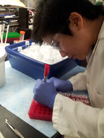
In the lab, there are many tools of the trade— one being pipettes. Pipettes are what we use to transfer liquids from one place to another. When putting reagents, medium, dna, etc. in many samples, you go through a lot of pipettes. Since each sample is different, you can't use the same pipette twice to avoid cross-contamination, leading to poor results. My mentor told me during the first week of internship that sometimes, instead of turning around and putting her used pipettes in the trash every time, she tries to stack them as high as she can. While I was mixing the GFP plasmid with the transfection reagent for my cells, I decided to try this out. Here, I managed to stack fifty used pipettes on top of each other. What I enjoyed about the lab and my mentor was that everyone would have little funny/fun things that they did making the work entertaining. What made this internship not only educational, but fun was the little things like this.
Transfecting the cells was actually the easy part. The process was not as hard as with other experiments I completed. Here I’m pipetting the GFP plasmid and the transfection reagent into my cells. The transfection reagent forms liposomes that trap the GFP inside of them, so the GFP can cross through the cell’s membrane. The transfection reagent then transports the GFP into the nucleus, where it is expressed. The problem with this is that there are a lot of factors that could interfere with this transportation system. The transfection reagent may never reach the nucleus, it could become trapped in the nuclear envelope if in a dividing cell, the transfection reagent may not be able to penetrate the nuclear membrane, the list goes on. To optimize the potential of the GFP reaching the nucleus, and being transcribed, I let the cells sit for forty-eight hours before checking. After this, I checked to see if the cells were properly transfected by running a western blot, and viewing to see if the cells fluoresce under a microscope.
|
|
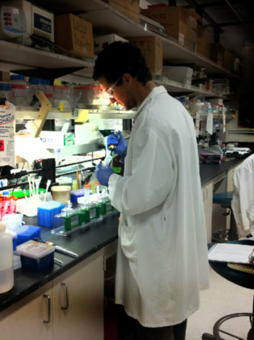
Western Blots are standard in labs. A western blot is when you separate proteins by the length of their polypeptides on a gel, by running an electric current through it. You can then transfer the proteins from the gel, onto a membrane by also using an electric current. Once on the membrane, you can see which proteins appeared the most in the cell. Since I transfected GFP in my cells, the GFP should be easily visible on the membrane when I run a western blot on it. You can see my western blot running in the photo on the left, then the protein on the membrane pictured as well. One other option here is to view the cells under a microscope, and excite them at the proper wavelength to see if they fluoresce. When I did this, the cells were fluorescing, so my transfection was successful.
|
